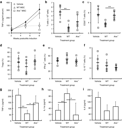
Figure 3.
Aire mediates suppression through T cell activation rather than effector T cell skewing or Treg induction. Colitis was induced and mice were treated as depicted in Figure 2a. Tissues were harvested 6 weeks postdisease induction. Mesenteric lymph nodes were isolated and cells activated in culture before analysis of intracellular cytokines. (a) Serum TNF-a levels were tested by ELISA following colitis induction. Flow cytometry of (b) mesenteric lymph node T cell numbers and (c) CD25+ T cells, shown as % total CD4+ T cells. (d) FoxP3+ Tregs, shown as % total CD4+CD25+ T cells. Intracellular cytokine flow cytometry of (e) IFN-g and (f) IL-10, shown as % total CD4+ T cells. Intestinal lesions were isolated during active colitis at 6 weeks after induction and cultured overnight in basal media. ELISA of (g) TNF-α, (h) IL-1ra, and (i) IL-6 release. Data depict mean ± SD and are representative of n = 3–5 per group from two experiments. *P < 0.05; **P < 0.01, Student's t-test. ELISA. Enzyme-linked immunosorbent assay; IFN, interferon; IL, interleukin; TNF, tumor necrosis factor; WT, wild type.

