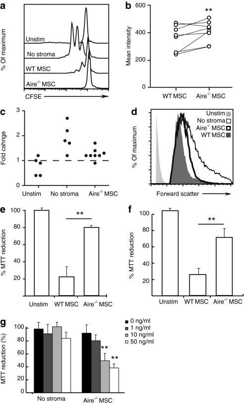
Figure 5.
Aire-regulated Eta-1 alters T cell size and mitochondrial reductase. T cells stimulated with anti-CD3ε were cocultured with WT or Aire−/− MSCs for 3 days. Unstimulated (Unstim) and cells stimulated but not cultured with stroma (“No stroma”) were used as negative and positive controls, respectively. (a) T cell proliferation was detected as CFSE dilution. (b) Mean forward scatter intensity for live T cells. (c) Fold change in live T cell mean forward scatter intensity, compared to T cells cocultured with WT MSCs (normalized to one). (d) Histograms showing forward scatter profiles gated on live CD4+ T cells that have undergone at least one cell division. (e) MTT reduction of CD3ε-activated T cells cocultured with WT or Aire−/− MSCs. (f) MTT reduction of CD3ε-activated T cells separated from MSCs by a porous membrane. (g) MTT reduction of CD3ε activated T cells, indirectly cocultured with Aire−/− MSCs, with increasing doses of Eta-1 neutralizing antibody. Data represent at least two independent experiments and three mice. (**P = 0.01, two-tailed Student's t-test; Aire−/− MSCs vs. WT (b,e,f) or 0 ng/ml antibody (g)).CFSE, carboxyfluorescein succinimidyl ester; MSC, mesenchymal stem cell; MTT, 3-(4,5-dimethylthiazol-2-yl)-2,5-diphenyl-tetrazolium bromide; WT, wild type.

