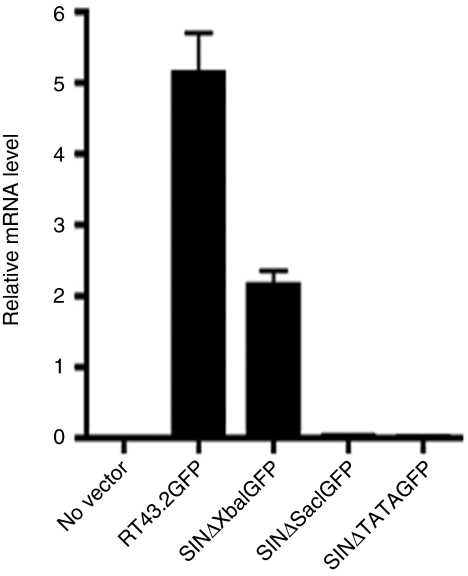
Figure 5.
Relative GFP mRNA levels in MDTF cells exposed to different MLV-based vectors. RNA was extracted from MDTF cells exposed to the indicated vectors. mRNA levels were determined by qRT-PCR. Relative GFP mRNA level was shown after normalization with GAPDH. Results are depicted as mean ± SEM, n = 3. GAPDH, glyceraldehyde 3-phosphate dehydrogenase; GFP, green fluorescent protein; MDTF, Mus dunni tail fibroblast; MLV, murine leukemia virus; mRNA, messenger RNA; qRT-PCR, quantitative reverse transcription-PCR.

