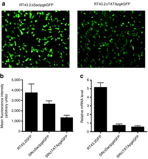
Figure 6.
An internal promoter, pgk, restored GFP expression in cells exposed to MLV-based vectors with larger deletions in the U3 region. (a) Representative fluorescence microscopic images of GFP expression in MDTF cells exposed to SINΔSacIpgkGFP vectors (left panel) or SINΔTATApgkGFP vectors (right panel). (b) MFI of MDTF cells exposed to RT43.2GFP, SINΔSacIpgkGFP or SINΔTATApgkGFP vectors as measured by flow cytometry. Results are depicted as mean ± SEM, n = 3. (c) Relative GFP mRNA level of MDTF cells exposed to RT43.2GFP, SINΔSacIpgkGFP or SINΔTATApgkGFP using qRT-PCR. Results are depicted as mean ± SEM, n = 3. GFP, green fluorescent protein; MDTF, Mus dunni tail fibroblast; MFI, mean fluorescence intensity; MLV, murine leukemia virus; mRNA, messenger RNA; qPT-PCR, quantitative reverse transcription-PCR; SIN vectors, self-inactivating vectors.

