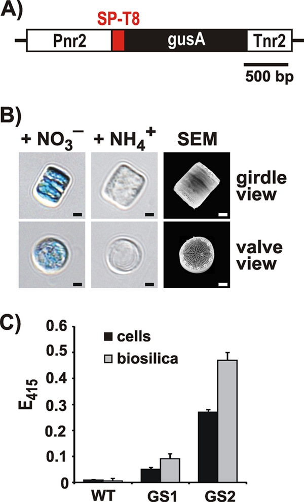
Fig 2.

Expression of the SP-T8-GUS fusion protein in T. pseudonana. (A) The recombinant fusion gene encoding SP-T8-GUS was placed under the control of the inducible expression cassette Pnr2/Tnr2 that was derived from the T. pseudonana nitrate reductase gene (30). (B) Histochemical assay for GUS activity (20) with cells from T. pseudonana transformant clones that contain the T8-GUS fusion protein. The cells were grown with ammonium (NH4+) or nitrate (NO3−) as the sole nitrogen source in the medium. The scanning electron micrograph (SEM) shows intact biosilica cell walls from two diatom cells, one in girdle view (top) and the other in valve view (bottom). Scale bars: 1 μm. (C) Photometric assay for GUS activity (20) with intact cells and isolated biosilica from wild-type (WT) T. pseudonana and two independent transformant clones (GS1, GS2). Equal numbers of cells from each strain were used for biosilica isolation and live cell activity assays. The height of each bar represents the average value from the analyses of three independent experiments.
