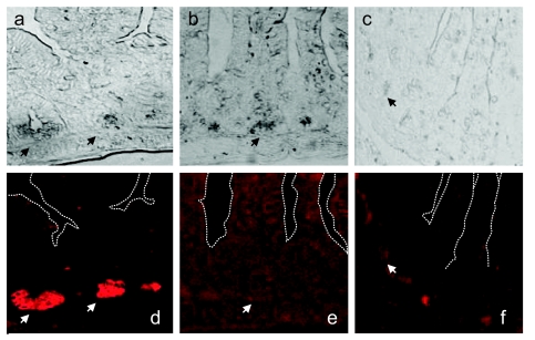
Fig 1.
Loss of P lysozyme-positive Paneth cells in ST-infected mice. Small-intestinal tissue sections collected 72 h after infection were stained with rabbit polyclonal antibodies against M and P lysozymes and Alexa 594-conjugated chicken anti-rabbit antibodies. The top row shows phase-contrast images. The bottom row shows images from a fluorescence microscope. (a and d) Tissue sections from mice inoculated with PBS; (b and e) tissue sections from same mice stained without primary antibody; (c and f) tissue sections from mice infected with ST. Images were taken at ×400 magnification. Arrows point to Paneth cells. The white dashed lines in panels d to f trace villus-crypt axes.

