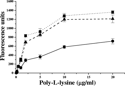
Fig 2.
Binding of FITC-labeled PLL to whole S. aureus cells. The graph shows the relative fluorescence units (± the SD) of FITC-labeled PLL bound to Mu50 (●), ΔvraG (■), and ΔgraR (▲) whole cells: the lower the number of fluorescence units, the greater the PLL repulsion and the more positively charged the S. aureus cell envelope (30).

