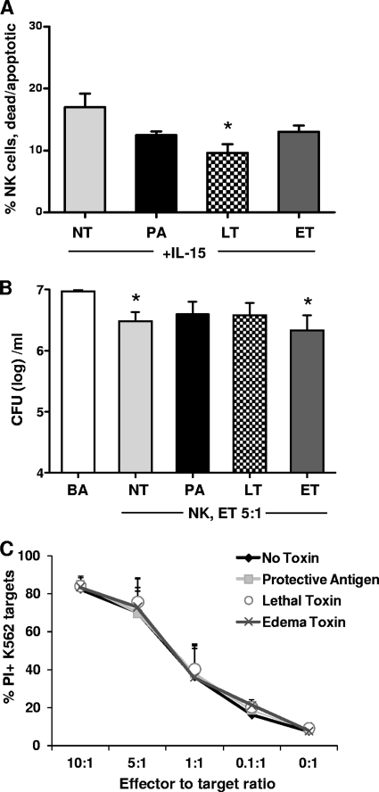
Fig 2.
Effects of B. anthracis LT and ET on NK cell antibacterial and cytotoxic function. Natural killer cells were isolated by use of a magnetic bead-conjugated antibody from PBMCs of healthy human donors and cultured with rIL-15 (15 ng/ml). (A) Flow cytometric analysis of NK cell viability, displayed as percent propidium iodide-positive annexin V-positive (dead/apoptotic) NK cells following 24 h of exposure to no toxin (NT), the protective antigen (PA) component, lethal toxin (LT), or edema toxin (ET). (B) CFU of B. anthracis (BA) bacilli following 24 h of in vitro coculture in media or with primary human NK cells (E/T ratio of 5:1) exposed to NT, PA, LT, or ET. (C) Cytotoxicity of NK cells treated with NT, PA, LT, or ET against the classical NK cell target K562 cells, displayed as percent PI-positive K562 cells. Values shown are the means ± SEM of results from 4 individual donors. *, P < 0.05 (P values indicate statistically significant differences due to treatment compared to NT [A] or B. anthracis [B]).

