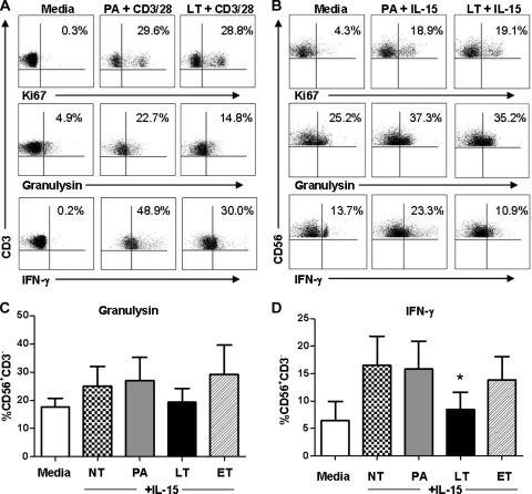
Fig 3.
B. anthracis LT suppresses activation of NK cell IFN-γ and T cell granulysin. Purified NK cells or PBMCs were rested (medium) or exposed to PA (negative control), LT, or ET and activated with IL-15 (15 ng/ml) or antibodies to CD3 and CD28. (A and B) Flow cytometric results of CD3+ CD56− (T cell; n = 4) (A) and CD56+ CD3− (NK cell; n = 6) (B) events from a representative donor. The results displayed are the effects of LT on the activation of T cell or NK cell proliferation (Ki67 proliferation marker) and the expression of intracellular granulysin and IFN-γ activated by IL-15 or CD3/CD28. (C and D) Summarized flow cytometry results for the expression of granulysin and IFN-γ by NK cells affected by exposure to LT or ET. Summarized values shown in panels C and D are the means ± SEM of results from 6 individual donors. *, P < 0.05 (P values indicate statistically significant differences due to treatment compared to the NT treatment).

