
Fig 6.
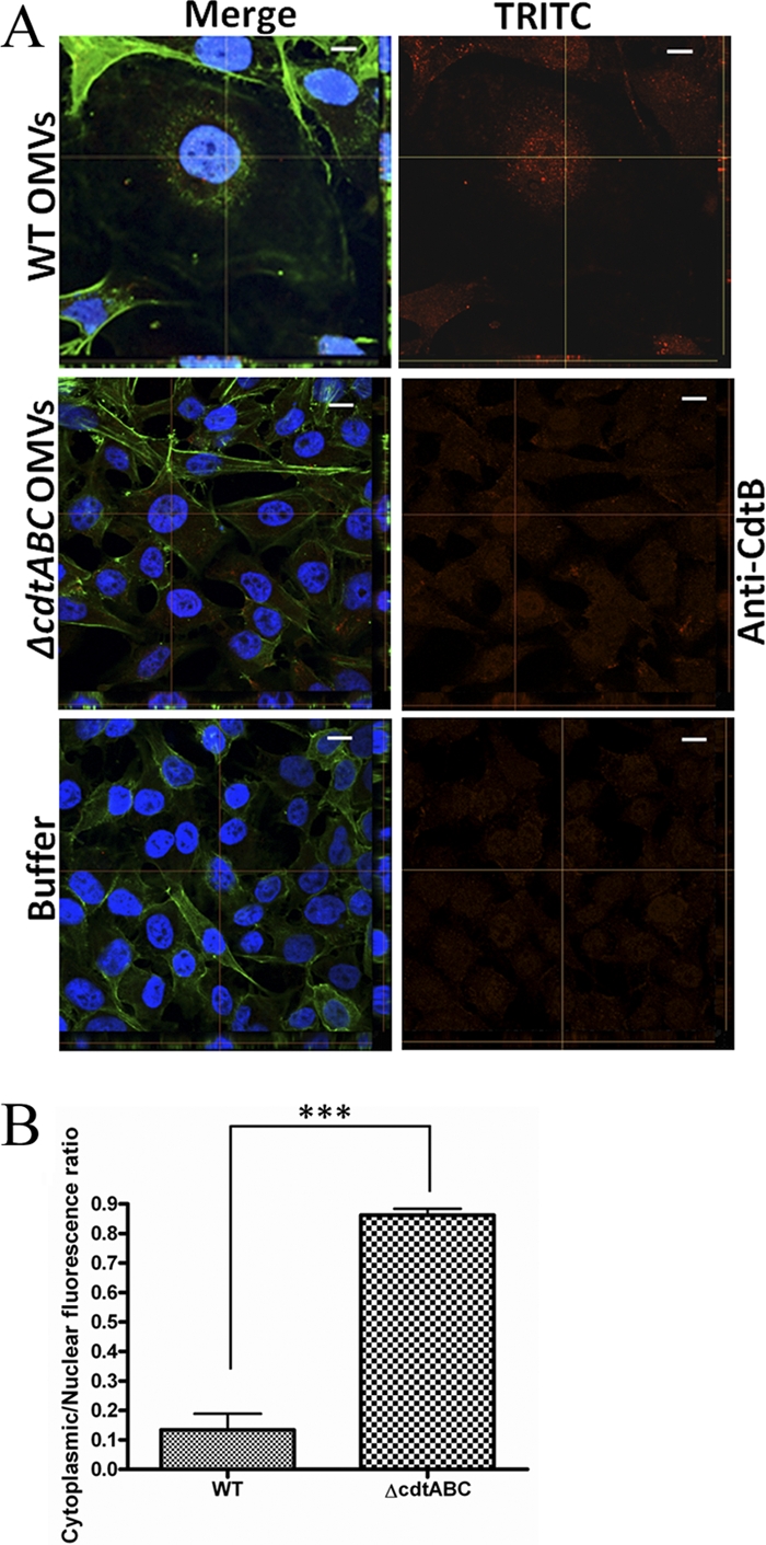
Nuclear distribution of internalized CdtB. (A) OMVs from A. actinomycetemcomitans strain D7SS (WT) or D7SS cdtABC (cdtABC) or PBS (buffer) were used to treat HeLa cells for 72 h. After treatment, cells were fixed and incubated with an antibody specific for H. ducreyi CdtB. Actin filaments and nuclei were then stained with phalloidin (green) and DAPI (blue), respectively. Bound antibodies were detected with TRITC (red). The left panels show merged images from staining with the three dyes. The right panels show images from TRITC staining only. Confocal Z-stack projections are included in all images. The cross hairs indicate the positions of the xz and yz planes. Magnification, ×1,000. Bars = 10 μm. (B) Assessment of the nuclear localization of CdtB. HeLa cells were treated with strain D7SS (WT) or D7SS cdtABC (cdtABC) OMVs for 72 h. Shown are the means ± SD for the cytoplasmic/nuclear ratio of red fluorescence from 20 cells from three experiments (P < 0.0001).