Fig 5.
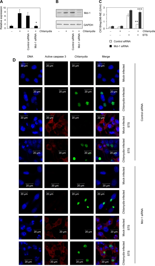
Silencing of Mcl-1 in IFN-γ-treated HeLa cells. Cells were transfected with specific or control siRNA for 48 h and then infected with C. trachomatis. IFN-γ stimulation was started at 48 h before infection. Cells were analyzed at 24 h after infection. (A) Downregulation of Mcl-1 mRNA levels was assessed by real-time RT-PCR. Data were normalized to GAPDH mRNA levels as described in Materials and Methods. *, P < 0.02 (compared to values for infected cells with or without control siRNA transfection). (B) Inhibition of Mcl-1 protein production following siRNA transfection. (C) Apoptotic cells in culture with and without STS treatment were determined by quantification of CK18Asp396-NE in cell lysates. **, P < 0.01 (compared to values for mock-infected cells transfected with control siRNA and exposed to STS); ***, P < 0.001 (compared to values for infected cells transfected with control siRNA and exposed to STS; n = 4). (D) Cell monolayers were costained with DAPI for DNA (blue), active caspase-3 antibody (probed with a Cy3-conjugated secondary antibody; red), and FITC-conjugated C. trachomatis MOMP antibody (green). For the experiments shown in panels C and D, 1 μM STS was added 3 h before sampling to induce apoptosis.

