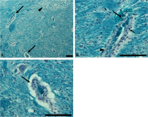
Fig 3.
Tissue from a piglet challenged with the positive-control Sakai strain exhibiting mortality on day 6 p.i. (A) Section of the brain stained with Luxol-fast blue stain showing affected blood vessel (arrows, magnified in panels B and C) with vacuolation of adjacent tissues (arrowhead) caused by focal myelin degeneration. (B) Affected blood vessel in brain showing microhemorrhage (long arrow), pyknosis (short arrow), and hyperplasia (arrowhead). (C) Affected blood vessel in brain showing perivascular edema. Bar = 100 μm.

