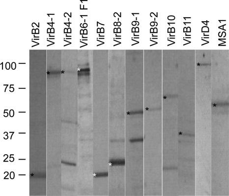
Fig 1.
Purity of recombinant T4SS proteins. Recombinant T4SS proteins were loaded at 1 μg/well, separated on 4 to 20% gradient SDS-PAGE gels, and stained with Coomassie blue dye. The proteins were electrophoresed on separate gels, and scanned images were arranged as presented. An asterisk indicates the predicted molecular mass of the recombinant protein, indicated in parentheses as follows: rVirB2 (20 kDa), rVirB4-1 (90 kDa), rVirB4-2 (91 kDa), rVirB6-1 F1 (90 kDa), rVirB7 (20 kDa), rVirB8-2 (23 kDa), rVirB9-1 (46 kDa), rVirB9-2 (46 kDa), rVirB10 (65 kDa), rVirB11 (38 kDa), rVirD4 (93 kDa), and control rMSA1 (58 kDa).

