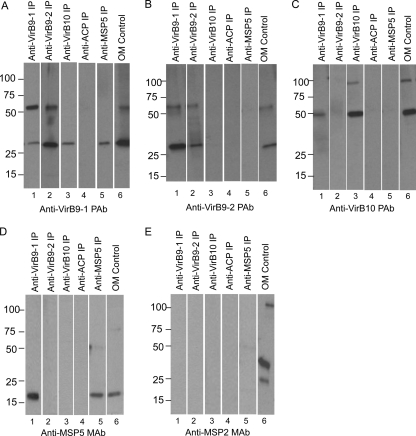
Fig 6.
Detection of native protein interactions of VirB9-1, VirB9-2, and VirB10. Solubilized A. marginale OM proteins were immunoprecipitated with purified rabbit IgG against VirB9-1 (lane 1), VirB9-2 (lane 2), VirB10 (lane 3), negative control B. bovis ACP (lane 4), and an MAb specific for MSP5 (lane 5) as indicated for each lane. Solubilized OM were included (lane 6) for a size comparison. Immunoprecipitated pellets and OM were electrophoresed and transferred to nitrocellulose membranes. Detection of the interacting proteins was performed by Western blotting individual strips with purified rabbit IgG against VirB9-1 (A), VirB9-2 (B), and VirB10 (C) and MAbs specific for MSP5 (D) and MSP2 (E), as indicated under each panel. The secondary antibody was Clear Blot-HRP (panels A to D and panel E, lane 5) or goat anti-mouse IgG+IgM (panel E, lanes 1 to 4 and 6). Scanned images were rearranged and presented as shown. The predicted molecular masses for native proteins are indicated in parentheses: VirB9-1 (30 kDa), VirB9-2 (30 kDa), VirB10 (49 kDa), B. bovis ACP (15 kDa), MSP5 (19 kDa), and MSP2 (37 kDa).

