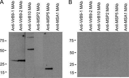
Fig 7.
MAb detection of interacting T4SS proteins immunoprecipitated by rabbit anti-VirB9-1 IgG. Solubilized OM proteins were immunoprecipitated with rabbit anti-ViB9-1 (A) or normal rabbit (B) IgG, and the pellets were electrophoresed and transferred to nitrocellulose membranes. Strips were cut and probed with MAbs specific for VirB9-1, VirB9-2, VirB10, MSP2, MSP5, and B. bovis MSA1, as indicated at the top of each lane. The secondary antibody was goat anti-mouse IgG+IgM. Scanned images were rearranged and are presented as shown. The predicted molecular masses for native proteins are indicated in parentheses: VirB9-1 (30 kDa), VirB9-2 (30 kDa), VirB10 (49 kDa), MSP2 (37 kDa), MSP5 (19 kDa), and B. bovis MSA1 (42 kDa).

