
Fig 3.
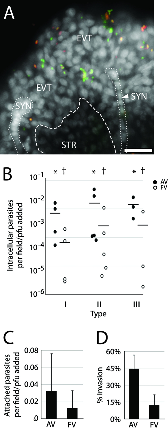
Comparison of T. gondii strains' invasion of placental explants. (A) Representative whole-mount placental explant infected for 5 h with type III T. gondii and stained with anti-SAG1 (red-stained parasites) without permeabilization. All living or recently dead parasites express GFP and are therefore green; extracellular parasites also stained red. This “inside-out” stain reveals parasites predominantly localized to EVT, SYN, or STR. White, DAPI. Bar, 100 μm. (B) Intracellular parasites were counted in AV and/or FV and normalized to the infectious dose as determined by PFU. Each dot represents the average of three fields per one placenta. * versus † denotes statistically similar groups (P < 0.04). Bars, SEM. (C) Average counts of extracellular attached parasites in each region (AV or FV) across all strains indicate a slight but not statistically significant pathogen preference for AV. (D) Counts of intracellular parasites as a fraction of total parasites found in AV and FV regions across all strains show invasion is significantly more likely (P > 0.0005) in AV. (C and D) Three AV or FV fields in each of three placentas per each of three strains. Bars, standard deviations (SD).