Fig 3.
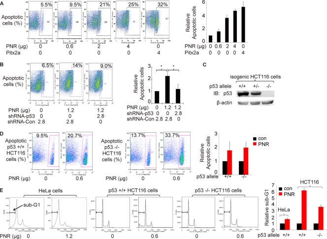
p53 enhances PNR-induced cell apoptosis. (A) PNR induces HeLa cell apoptosis in a dose-dependent fashion. Two days after transfection, an annexin V binding assay was used to quantitate apoptotic cells by flow cytometry. Pitx2a was used as a positive control (50). The fraction of apoptotic cells in the sample with 0 μg of PNR was normalized to 1. Data representing the results of three repeated experiments are plotted on the right. (B) shRNAs targeting p53 significantly inhibit PNR-induced HeLa cell apoptosis. shRNA-Con, pLKO.1 empty expression vector control. The right plot shows relative levels of apoptosis based on the means of data determined under each set of conditions in 3 independent experiments. The mean for samples with only shRNA-Con was normalized to 1. *, P < 0.05 (one-tailed test). (C) p53 protein levels in three isogenic HCT116 cell lines containing two p53 alleles, one allele, or none. (D and E) PNR induces cell apoptosis in both p53+/+ and p53−/− HCT116 cells. Two days after transfection of PNR expression plasmid, the isogenic HCT116 cells were processed for both an annexin V binding assay and sub-G1 analysis. The data from three repeated experiments is plotted on the right. (D) Annexin V binding assay. PNR mildly induced apoptosis at similar levels in both cell lines. (E) Sub-G1 analysis. PNR induced apoptosis in p53+/+ HCT116 cells at a level ∼2-fold higher than the level seen in p53−/− HCT116 cells. PNR also increased apoptosis at the sub-G1 phase in HeLa cells.

