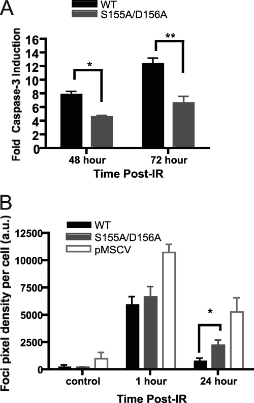
Fig 4.
Ku70 S155A/D156A-expressing cells exhibit DNA damage signaling defects. (A) Analysis of IR-induced apoptosis in Ku70 S155A/D156A-expressing cells. Irradiated or mock-treated Ku70−/− MEFs expressing wild-type Ku70 (WT) or Ku70 S155A/D156A were assayed for caspase-3 activity at the times indicated. The fold activation of caspase-3 activity is shown relative to that of the unirradiated control and averaged for four experiments, with error bars representing the SEM (∗∗, P < 0.01; ∗, P < 0.05). (B) S155A/D156A Ku70 mutant cells display prolonged H2AX serine 139 phosphorylation (γ-H2AX) 24 h after IR. As described for panel A, cells were irradiated with 4 Gy of IR or mock treated, fixed at the time points indicated, and subjected to analysis with a γ-H2AX antibody and DAPI. Foci were quantified based on pixel intensity and averaged for the number of cells present (a.u., arbitrary units). Data represent averages from four separate experiments, each assessing approximately 500 cells, and error bars represent SEM (∗, P < 0.05).

