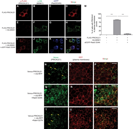
Fig 8.
Rab5 is involved in PRICKLE1 trafficking. FLAG-PRICKLE1 was expressed alone (A to D), in the presence of HA-MINK1 (E to H), or in the presence of HA-MINK1 and a dominant-negative version of Rab5, eGFP-Rab5 (S34N) (I to L). Expressed alone, PRICKLE1 is localized in the cytoplasm and evenly at the plasma membrane (A); when MINK1 is expressed, PRICKLE1 is redistributed within plasma membrane patches (E), whereas this membrane accumulation is inhibited by the overexpression of a dominant-negative mutant of Rab5 (I). Scale bars represent 20 μm for panels A to L. (M) The quantification of PRICKLE1-containing plasma membrane patches in cells expressing PRICKLE1 alone, PRICKLE1, and MINK1 or within cells coexpressing PRICKLE1, MINK1, and Rab5 (S34N). Error bars indicate ±SEM. Statistical analysis was performed using one-way ANOVA followed by Tukey postanalysis (***, P < 0.001). In the absence of MINK1, PRICKLE1 was never present in plasma membrane patches (n = 83). The overexpression of MINK1 led to 89% ± 5% (n = 244) of the cells displaying punctum accumulation of PRICKLE1. This patchy localization of PRICKLE1 induced by MINK1 was inhibited by the overexpression of the dominant-negative mutant of Rab5, with 6% ± 4% (n = 196) harboring these structures. The images and the quantification shown are representative and tallied from three independent experiments. The punctum localization of Pk during Xenopus CE requires Rab5. Venus-PRICKLE1 mRNA (500 pg) and membrane-bound RFP (mb-RFP) mRNA (100 pg) were injected alone (N to P) or together with 1 ng of dominant-negative Rab5 (S34N) (Q to S) or Rab4 (S27N) (T to V) in dorsal blastomeres of 4-cell Xenopus embryos. Pk cellular localization was monitored in DMZ explants at stage 11. (T to V) In the presence of dominant-negative Rab4 (S27N), PRICKLE1 localization is not affected. Yellow arrows indicate plasma membrane puncta containing Venus-Pk. (Q to S) The coexpression of Rab5 (S34N) inhibits the localized accumulation of PRICKLE1 at the plasma membrane. Scale bars represent 50 μm in panels N to V.

