Fig 5.
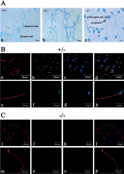
Acrosome and axoneme in Odf1-mutant sperm. (A) Acrosome reaction in epididymal sperm of wild-type (+/+), heterozygous (+/−), and homozygous (−/−) mice. A characteristic light blue staining of the reacted acrosome is visible in wild-type (a; +/+) and heterozygous (b; +/−) sperm, whereas in Odf1−/− sperm (c; −/−) no acrosome reaction was found. In addition, a remarkable sperm coiling in Odf1−/− sperm is visible. Bars: 10 μm. (B and C) Detection of the axonemal microtubules in heterozygous (B) and homozygous (C) Odf1-deficient spermatozoa. Sperm were released from the cauda epididymides of heterozygous (B, +/−) and homozygous (C, −/−) mice and stained for the acrosome with PL-FITC (in green) and for tubulin with anti-α-tubulin antibodies (in red). Nuclear counterstain with DAPI is shown in panels c, g, k, and o (in blue). In heterozygous and homozygous sperm the sperm tail is decorated with anti-α-tubulin antibodies demonstrating the presence of the axonemal structure (a, e, i, and m). In homozygous sperm the tail showed a conspicuous coiling (i, m, l, and p). The acrosome is present in heterozygous sperm (b and f) but absent in homozygous sperm (j and n). In addition, characteristic DAPI-stained sperm heads are also barely visible in homozygous preparations (k and o).

