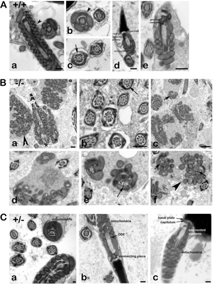
Fig 6.
Ultrastructure of spermatozoa. (A) Transmission electron microscopy of epididymal spermatozoa from wild-type mice. The well-structured organization of the sperm is shown, including the mitochondrial sheath (a, b, d, and e, arrowheads), the outer dense fibers (c; arrow), and the neck region (d and e). Bars: 250 nm (a and d), 500 nm (e). (B) Transmission electron microscopy of Odf1−/− sperm. Epididymides (a to c) and testes (d to f) from Odf1-deficient mice were prepared, and sections analyzed by electron microscopy. Sperm are highly disorganized including the mitochondrial sheath (a, c to f, arrowheads), as well as the outer dense fibers in the midpiece (c to f, arrows) and in the principal piece (b, arrow). In addition, very often in the cytoplasm of one cell more than one axonemal section was found (two longitudinal sections of axonemata in panel a, asterisks). Arrowheads, disturbed mitochondrial sheath; arrows, disturbed alignment of ODFs. Bars: 500 nm (a and b), 1,000 nm (c). (C) Transmission electron microscopy of sperm from heterozygous animals. No obvious disturbances are found but instead well-organized mitochondria (a to c), ODF (a and b), and connecting piece (b and c). Bars: 100 nm (a), 250 nm (b and c).

