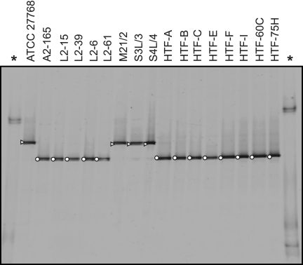
Fig 3.
PCR-DGGE fingerprints from F. prausnitzii isolates. Isolates are distributed in two separate bands that correlate with phylogroup designation (▵, phylogroup I; ○, phylogroup II). Asterisks indicate the ladder lanes (made by 16S rRNA gene fragments of Mucor sp., Pseudomonas fluorescens, and Micrococcus luteus, respectively, from the top to the bottom).

