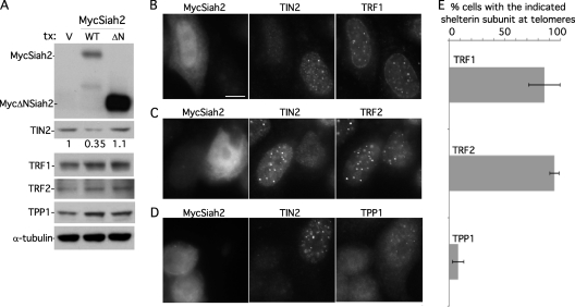
Fig 6.
TRF1 and TRF2, but not TPP1, remain at telomeres upon Siah2-mediated loss of TIN2. (A) Immunoblot analysis shows that MycSiah2 (but not MycΔNSiah2) induces loss of TIN2 protein but not TRF1, TRF2, or TPP1. Results of immunoblot analysis of extracts from supertelomerase HeLa cells transfected with vector (V), MycSiah2 (WT), or MycΔNSiah2 (ΔN) and probed with anti-TIN2 701, anti-TRF1 415, anti-TRF2, anti-TPP1 911, or anti-α-tubulin antibodies are shown. TIN2 protein levels relative to α-tubulin and normalized to the vector control are indicated below the upper blot. (B to D) Immunofluorescence analysis of supertelomerase HeLa cells transfected with MycSiah2. Cells were formaldehyde fixed and triply stained with anti-myc, anti-TIN2 701, and anti-TRF1 (B), anti-TRF2 (C), or anti-TPP1 1146 (D) and DAPI. Bar, 5 μm. (E) Graphical representation of the frequency of Siah2-expressing TIN2-lacking cells with the indicated shelterin component at telomeres. Data represent the means ± standard deviations of the results of three independent experiments (n = 69 cells or more each).

