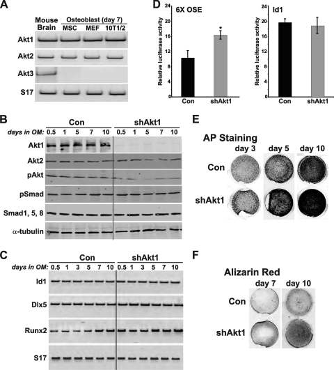
Fig 1.
Akt1 deficiency enhances BMP2-stimulated osteoblast differentiation. (A) Detection by reverse transcription-PCR (RT-PCR) of mRNAs for Akt1, Akt2, Akt3, and S17 in RNA from mouse brain (positive control), and from osteoblasts derived from MSCs, MEFs, and C3H10T1/2 mesenchymal stem cells after 7 days of differentiation. (B to F) C3H10T1/2 cells, infected with Ad-shAkt1 or Ad-βgal (Con), were incubated in osteogenic medium (OM) with BMP2 (200 ng/ml) for up to 15 days. (B) Immunoblots of whole-cell protein lysates for Akt1, Akt2, phospho-Akt (pAkt), pSmad, Smad1, 5, 8, and α-tubulin. (C) Measurement of mRNAs for Id1, Dlx5, Runx2, and S17 by RT-PCR. (D) Results of luciferase activity assays after transient transfection with reporter plasmids containing six copies of a Runx2 responsive element from the osteocalcin gene (OSE) or the BMP2-responsive mouse Id1 promoter and incubation in OM with BMP2 for 2 days (mean ± standard error of results of three experiments, each performed in triplicate; ∗, P < 0.01 versus Con). (E) Alkaline phosphatase (AP) staining on days 3, 5, and 10. F. Alizarin red staining for mineralization on days 7 and 10.

