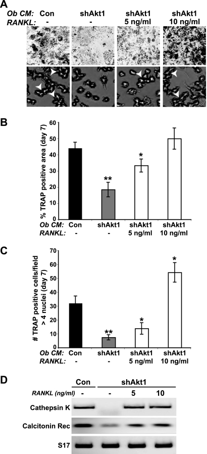
Fig 10.
RANKL restores osteoclast differentiation when added to conditioned medium from osteoblasts lacking Akt1. Conditioned medium (Ob CM) was collected from control (Con) C3H10T1/2 cells or cells infected with Ad-shAkt1 after incubation in osteoblast differentiation medium plus BMP2 for 7 days and was added to mouse bone marrow macrophages (as described in the legend to Fig. 5) without or with recombinant mouse RANKL (5 or 10 ng/ml). (A) Images of TRAP staining to assess formation of multinucleated osteoclasts on day 7 (top row, ×40 magnification; bottom row, ×200 magnification; white arrowheads indicate osteoclasts with >4 nuclei). (B) Percentage of TRAP-positive osteoclasts on day 7 expressed as area per microscopic field (×200 magnification; mean ± SD of results of six fields; ∗, P = 0.01; ∗∗, P < 0.002 versus Con CM). (C) Percentage of multinucleated TRAP-positive osteoclasts on day 7 per microscopic field (n > 4 nuclei; ×200 magnification; mean ± SD of results of three fields; ∗, P = 0.02; ∗∗, P = 0.002 versus Con CM). (D) Expression by RT-PCR of mRNAs for cathepsin K, the calcitonin receptor (Rec), and S17 on day 7.

