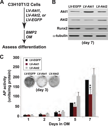
Fig 4.
Forced overexpression of Akt1 but not Akt2 decreases BMP2-mediated osteogenic differentiation. (A) Experimental scheme: C3H10T1/2 cells were infected with LV-Akt1, LV-Akt2, or LV-EGFP and were incubated in OM with 200 ng/ml BMP2 for up to 7 days. (B) Immunoblots for Akt1, Akt2, Runx2, and α-tubulin on day 7. (C) Quantitative measurement of AP activity on days 1 to 7 (mean ± SD of results of three experiments; ∗, P < 0.05; ∗∗, P < 0.01 versus cells infected with LV-EGFP). Inset shows AP staining on day 3.

