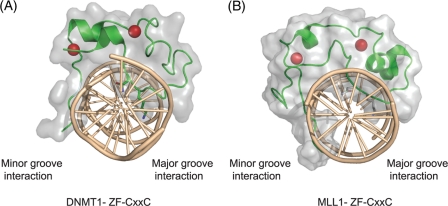
Fig 2.
ZF-CXXC domain proteins bound to nonmethylated CpG DNA. (A) A cartoon depicting the structure of the DNMT1 ZF-CXXC domain bound to nonmethylated CpG DNA (Protein Data Bank [PDB] number, 3PT6). The green ribbon indicates the protein backbone and the residues that specifically interact with CpG dinucleotides in the major groove. The DNA is colored light brown and is viewed looking down the DNA double helix. The gray shading indicates the protein surface, and the red spheres correspond to the two zinc ions coordinated by the ZF-CXXC domain. (B) The same representation as in panel A of the MLL1 ZF-CXXC domain bound to nonmethylated CpG DNA (PDB number, 2KKF). From both panels it is apparent that the ZF-CXXC domain interacts specifically with CpG DNA in the major groove and interacts with minor-groove DNA on the opposite side of the DNA double helix.

