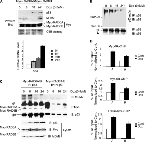
Fig 5.
RAD6 functions in the process of stress stimulus-induced p53 upregulation. (A) Doxorubicin (Dox) treatment causes the upregulation of p53 in HeLa cells. HeLa cells were transfected with the Myc-RAD6 plasmid and treated with doxorubicin (0.5 μM) for the indicated times. The immunoblot was stained with anti-p53, anti-MDM2, or anti-Myc antibodies. The histogram at the bottom shows the real-time RT-PCR analysis of the transcriptional levels of p53. (B) Doxorubicin (Dox) treatment inhibits the ubiquitination of p53 in HeLa cells. HeLa cells were treated with doxorubicin (0.5 μM) for different lengths of time. Coimmunoprecipitation analysis was employed using an anti-p53 antibody. The anti-p53 antibody was also used to visualize the amounts of precipitated p53 (lower panel) and ubiquitinated p53 (upper panel). (C) Doxorubicin (Dox) treatment inhibits the formation of the RAD6-MDM2-p53 degradation complex. HeLa cells were transfected with the Myc-RAD6 plasmid and treated with doxorubicin (0.5 μM) for the indicated periods of time. A coimmunoprecipitation analysis was employed using an anti-p53 antibody. The anti-p53, anti-MDM2, and anti-Myc antibodies were used to visualize the amounts of precipitated p53, MDM2, and RAD6, respectively (upper panels). The gel on the right shows an identical control IP using normal mouse IgG (NIgG). The expression levels of p53, MDM2, and RAD6 are shown in the lower panels. (D) Doxorubicin (Dox) treatment promotes the recruitment of RAD6 and H3K4me3 to the promoter (bars A) and 5′ coding (bars B) regions of the p53 gene. HeLa cells were transfected with the Myc-RAD6 plasmid and treated with or without (Cont.) doxorubicin (0.5 μM) for 24 h. The ChIP-qPCR analysis showed that doxorubicin treatment resulted in increased levels of RAD6 and H3K4me3 at these regions of the p53 gene.

