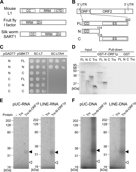
Fig 1.
ZfL2-1 ORF1p interacts with itself and binds to nucleic acids through the N-terminal region. (A) Schematic diagrams of LINE ORF1ps biochemically studied so far. The domain structures were predicted by HHpred (30). CC, coiled-coil motif; RRM, RNA recognition motif; CTD, C-terminal domain; Zn, zinc knuckle motif. (B) Schematic diagram of the zebrafish LINE, ZfL2-1. ZfL2-1 is composed of a 5′ untranslated region (UTR), two open reading frames (ORF1 and ORF2), and a 3′ UTR. The full-length ZfL2-1 ORF1p (FL) and its N- and C-terminal portions (N and C) were used in this study. Numbers above the boxes indicate the amino acid positions from the first methionine. ES, esterase domain. (C) Yeast two-hybrid assay. The pGADT7 derivatives express the activation domain of GAL4 fused with the N- or C-terminal portion of ZfL2-1 ORF1p. The pGBKT7 derivatives express the binding domain of GAL4 fused with the full-length ORF1p or its N- or C-terminal portion. AH109 cells that have the two derivatives were spotted in 10-fold serial dilutions on synthetic complete medium depleted of leucine and tryptophan (SC-LT) or that depleted of leucine, tryptophan, adenine, and histidine (SC-LTAH) and grown at 30°C for 3 days. (D) Autoradiogram of GST pulldown assay. Trx and the Trx fusions of the full-length, N-terminal portion, and C-terminal portion of ZfL2-1 ORF1p were synthesized in vitro as 35S-labeled proteins and incubated with the GST fusion of full-length ZfL2-1 ORF1p (GST-F-ORF1p) or GST. Equal amounts of the 35S-labeled proteins before the incubation were subjected to SDS-PAGE (Input). 35S-labeled proteins bound to GST-F-ORF1p or GST were purified and electrophoresed on the gel (Pull-down). Sizes of standard marker proteins are indicated at the left of the autoradiogram. (E and F) UV cross-linking assays. Trx or the Trx fusion of the N-terminal portion of ZfL2-1 ORF1p (Trx-N-ORF1p) was incubated with 32P-labeled RNA (E) derived from the pUC19 vector (pUC-RNA) or the 3′ tail of ZfL2-1 (LINE-RNA) and 32P-labeled DNAs (F) derived from the pUC19 vector (pUC-DNA) or the 3′ tail of ZfL2-1 (LINE-DNA). Black and white arrowheads indicate the positions of Trx-N-ORF1p and Trx, respectively. The left lane of each autoradiogram shows the results with no protein (−). Sizes of standard marker proteins are indicated at the left of each autoradiogram. The RNA and DNA sequences are shown in Table 1.

