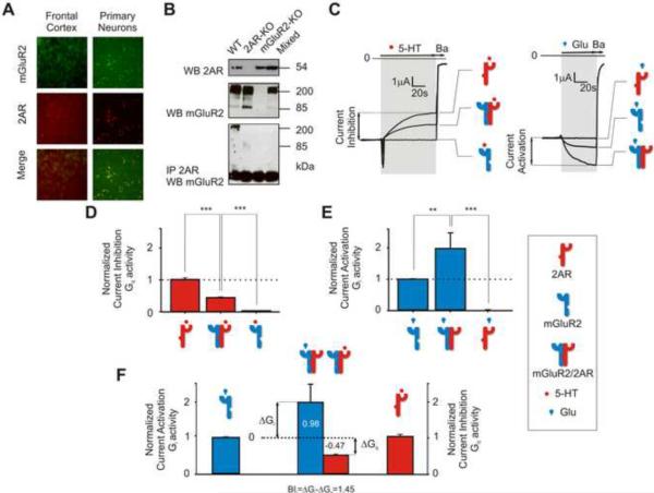
Figure 1. Heteromeric Assembly of 2AR and mGluR2 enhances Glu-induced Gi Signaling and Reduces 5-HT-induced Gq Signaling.
2AR and mGlu2 co-localize and form a receptor complex in mouse frontal cortex. (A) Representative micrographs showing co-expression of endogenous 2AR (red) and mGluR2 (green) in mouse frontal cortex (left panels) and mouse cortical primary neurons (right panels). (Scale bar 25 μm). (See also Figure S1 E)
(B) Mouse frontal cortex membrane preparations were immunoprecipitated (IP) with anti-2AR antibody. Immunoprecipitates were analyzed by western blot (WB) with anti-mGluR2 antibody (lower blot). Mouse frontal cortex membrane preparations were also directly analyzed by WB with anti- 2AR antibody (upper blot) or anti-mGluR2 antibody (middle blot). 2AR-KO and mGluR2-KO mouse frontal cortex tissue samples were processed identically and used as negative controls. Frontal cortex tissue samples from 2AR-KO and mGluR2-KO mice were also homogenized together (mixed) and processed identically for immunoprecipitation and WB.
(C) (Left) Representative barium-sensitive traces of IRK3 currents obtained in response to 1 μM 5-HT in oocytes expressing 2AR alone, mGluR2 and 2AR together, or mGluR2 alone. (Right) Representative barium-sensitive traces of GIRK4* currents obtained in response to 1 μM Glu in oocytes expressing mGluR2 alone, mGluR2 and 2AR together, or 2AR alone. Barium (Ba) inhibited IRK3 and GIRK4* currents and allowed for subtraction of IRK3 and GIRK4*-independent currents. For illustrative purposes, traces with similar basal currents were chosen.
(D) Summary bar graphs of Gq activity measured as IRK3 current inhibition (mean ± SEM) following stimulation with 5-HT and (E), of Gi activity measured as GIRK4* current activation (mean ± SEM) following stimulation with Glu. IRK3 current inhibition was measured relative to basal currents and was normalized relative to that obtained by stimulating 2AR alone with 5-HT (100% or 1). GIRK4* current activation was measured relative to the basal currents and was normalized relative to that obtained by stimulating mGluR2 alone with Glu (100% or 1) (F) Calculation of the balance index (BI) as the difference of the increase in Gi-signaling in response to Glu from the mGluR2 homomeric level (ΔGi) and the decrease of Gq-signaling in response to 5-HT from the 2AR homomeric level (ΔGq). A reference BI (Bir=1.45) was calculated for the mGluR2/2AR complex in response to 1 μM Glu and 1 μM 5-HT using mean values (** p<0.01, *** p < 0.001). See also Figures S1, S2, and S3.

