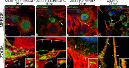
Fig 2.
Alignment and colocalization of EGFP-tagged or immunolabeled capsid proteins with microtubules in virus-infected TN-368 cells. TN-368 cells infected with individual recombinant AcMNPV expressing EGFP fusion (green) as EGFP-Orf1629 (a, a′, and a″), EGFP-EXON0 (b, b′, and b″), or EGFP-VP39 (c, c′, and c″) or infected AcMNPV and the VP39 immunolabeled (d, d′, and d″) using mouse α-VP39 antibody/Alexa Fluor 488 goat anti-mouse IgG (green). Cells were stained for microtubules with anti-α-tubulin/Alexa Fluor 568 goat anti-mouse IgG (red) and DNA with Vectashield DAPI (4′,6-diamidino-2-phenylindole; blue). Merged channels are shown in panels a, b, c, and d; zoomed sections of merged channels are shown in panels a′, b′, c′, and d′ and insets a″, b″, c″, and d″. White solid arrows indicate probably nucleocapsids. Bar, 10 μm.

