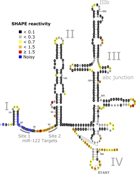
Fig 1.
Secondary structure of the HCV 5′-UTR. The 5′-UTR consists of 4 domains, labeled I to IV. The proposed secondary structure is consistent with all available crystallographic and/or NMR structural data (see Fig. S2a in the supplemental material). Colors denote SHAPE reactivity, as indicated, which is proportional to the probability that a nucleotide is single stranded.

