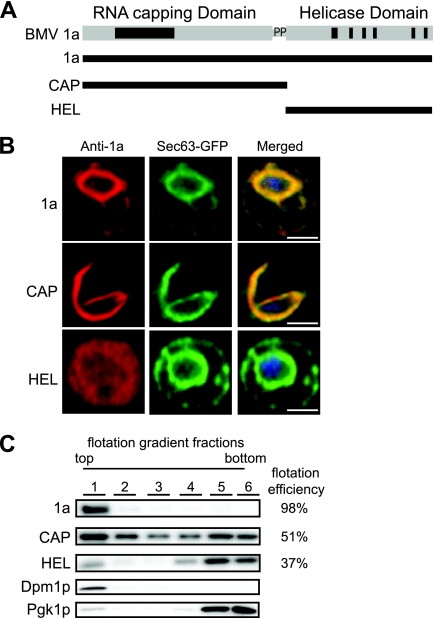
Fig 1.
CAP is mainly responsible for 1a membrane association. (A) The CAP fragment contains the capping domain and the proline-rich linker region, and the HEL fragment contains the NTPase/helicase-like domain. (B) Fluorescence microscopy images of cells expressing wt 1a, CAP, or HEL and Sec63-GFP, an ER marker. TO-PRO-3 was used to stain DNA (blue). Bars, 2 μm. (C) Distribution of 1a, CAP, HEL, PGK (cytosolic protein control), and Dpm1p (ER luminal protein control) in membrane flotation gradients. Representative Western blots using anti-1a, anti-PGK, and anti-Dpm1p antisera are shown.

