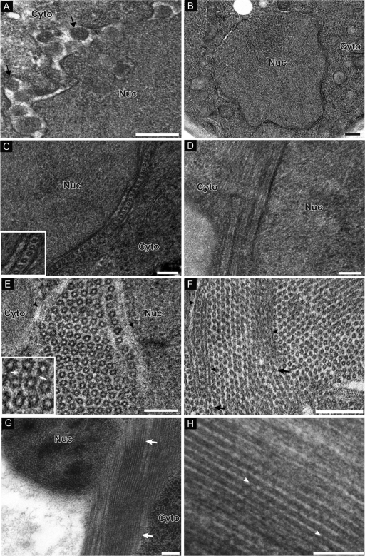
Fig 2.
CAP fragment induces formation of ring/tubular structures. (A) Electron micrograph of yeast cells expressing 1a-induced spherules. Arrows point out individual spherular structures. (B) HEL does not induce any membrane rearrangements. (C) Micrograph of yeast cells expressing CAP. In one subset of cells, nuclei were surrounded by appressed layers of double-membrane ER. When cross-sections were viewed, hexameric ring-like structures were present in the intermembrane spaces. The inset shows a close-up view of rings. (D) In views of longitudinal section, there are lamina-like structures present between membrane bilayers. (E and F) Alternatively, hexagonal lattices that either were not immediately flanked by an ER membrane (E) or contained ER-like layers a few hundred nanometers in length isolating each side of a single row of rings (F) were present. In the inset in panel E, arrows point to filamentous material connecting adjacent rings. Arrowheads in panel F point out membrane protrusions within the hexagonal ring lattice. (G) Another subset of cells displayed long electron-dense tubular structures partially surrounding the nucleus. (H) Close-up view of tubules. Arrowheads point out electron-dense material connecting tubules running parallel to each other. Nuc, nucleus; Cyto, cytoplasm. Bars, 200 nm (A, B, C, and H) and 100 nm (D to G).

