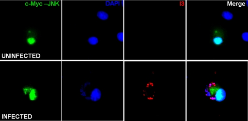
Fig 2.
Phospho-JNK localization during VACV infection. BSC-40 cells were transfected with pcDNA3-cMyc-JNK2-MKK7 for 48 h, followed by infection with VACV for 7 h. At this time, cells were processed for fluorescence analyses and simultaneously stained with anti-c-Myc and antiviral protein I3 antibodies, followed by FITC-conjugated antimouse and rhodamine-conjugated antirabbit antibodies (green and red, respectively). To detect cellular and viral DNA, cells were stained with DAPI (blue).

