Fig 3.
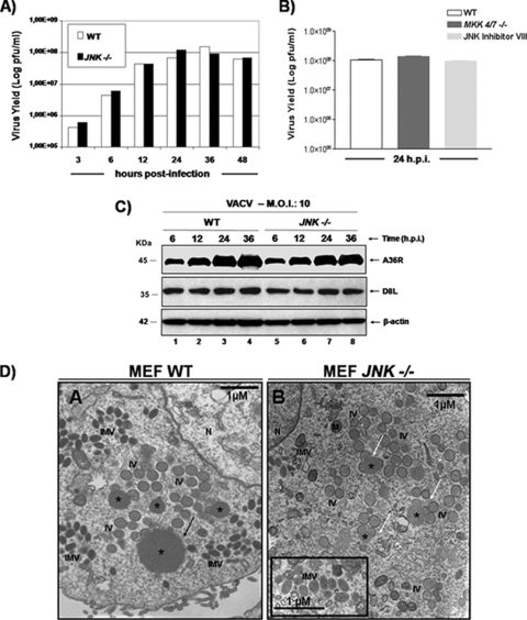
The VACV-stimulated MKK4/7-JNK1/2 pathway is not required for virus replication. (A) Growth curve of VACV performed in WT and JNK1/2-KO MEFs. Cells were infected with VACV at an MOI of 10 and harvested at 3, 6, 12, 24, 36, or 48 hpi, and viral yield was quantitated by viral plaque assay. Data are representative of at least three independent experiments. (B) WT MEFs were incubated in either the absence or presence of JNKi (4 μM) for 30 min. WT and MKK4/7-KO cells were infected (MOI, 10) with VACV for 24 h and assayed for viral production. Data are means of triplicate experiments + SDs. (C) WT or JNK1/2−/− MEFs were infected (MOI, 10) with VACV for the times shown, and 40 μg of WCEs was subjected to Western blotting. (Top and middle) Immunoblots with anti-A36R or anti-D8L antibodies; (bottom) anti-β-actin antibody was used as a loading control. The molecular masses (kDa) are indicated on the left. Data are representative of three independent experiments with similar results. (D) Confluent monolayers of WT and JNK1/2−/− MEFs were serum starved for 12 h and then infected with VACV at an MOI of 2 for 18 h. Cells were fixed and prepared for transmission electron microscopy. Micrographs are shown with their scale indicated by the bars. WT and JNK1/2−/− MEFs contain all the normal intermediates in virion morphogenesis (A and B). (B, inset) Detail highlighting the IMV form. Abbreviations: *, virosomes; IV, immature virions; IMV, intracellular mature virions; M, mitochondria; N, nucleus.

