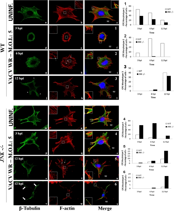
Fig 4.
VACV-infected JNK1/2−/− MEFS exhibit reduced early cell contractility. WT (a to l) and JNK1/2-KO (m to x) MEFs were infected with VACV WR (MOI, 5). At 3, 6, or 12 hpi, cells were fixed and stained for the microtubule and actin networks. β-Tubulin, F-actin, and the nucleus are stained with FITC-conjugated antimouse secondary antibody (green), rhodamine conjugated-phalloidin (red), and DAPI (blue), respectively. Fluorescently labeled cells were visualized using a Zeiss (LSM 510 META) confocal microscope. The graphic representations of the relative abundance of each phenotype found in WT-infected cells (graphs 1 to 3, f, i, and l) or JNK1/2−/−-infected cells (graphs 4 to 6, r, u, and x) are shown on the right (n > 100). (Left column) Thin arrows, microtubule projections (j); large arrows, microtubule long protrusions (v); (middle column) insets, cortical F-actin in detail, highlighting the presence (b, h, n, q, and t) or absence (e, k, and w) of stress fibers; dashed arrows, actin tails (h and t); (right column) insets, the edge of cell protrusions in detail, highlighting the protraction (c, i, l, o, r, u, and x) or retraction (f) of microtubule. Insets represent enlargement of the area indicated by the white box in the panel.

