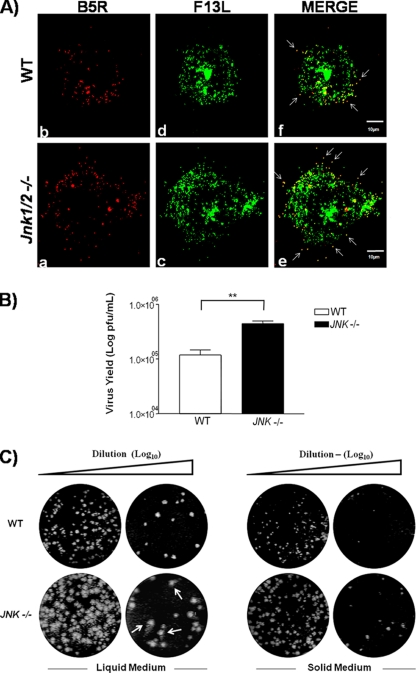
Fig 7.
Viral trafficking to the cell periphery is deregulated in JNK1/2-KO cells. (A) Detection of CEV in the cell surface by confocal microscopy. WT and JNK1/2-KO MEFs were infected with VACV vF13L-GFP at an MOI of 5 for 9 h. Cells were fixed and blocked without permeabilization and then incubated with mouse anti-B5 antibody, followed by staining with rhodamine-conjugated antimouse secondary antibody. White arrows indicate CEVs. Micrographs are shown with their scales indicated by the bars. (B) Absence of JNK1/2 affects the release of EEV from the infected cells. Culture supernatants of VACV-infected WT and JNK1/2/KO cells at an MOI of 10 for 48 h were incubated with the anti-IMV-specific L1R antibody (1:1,000) at 37°C for 1 h and then titrated in BSC-40 cells. Data are representative of at least three independent experiments with similar results (**, P < 0.05). (C) Viral plaque size is increased in the KO cells. WT and JNK1/2-KO MEFs were infected with serial dilutions of VACV in liquid or agarose-supplemented (solid) medium. At 72 hpi, cells were fixed and stained with crystal violet. Comet-like plaques (arrows) were visualized in the KO cells.

