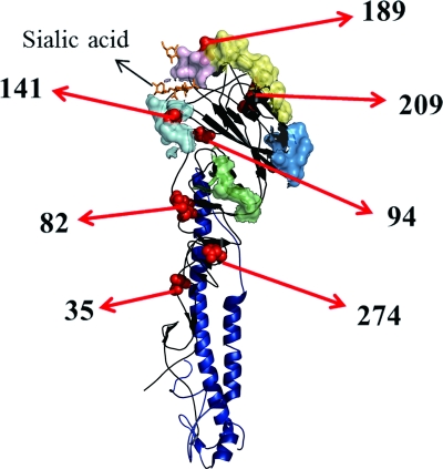
Fig 3.
Structure of HA monomer showing antigenic sites and amino acid residues at which substitutions occurred between 2004 and 2008. The drawing shows the HA monomer of the 1918 H1N1 virus, modified from the Protein Data Bank structure 2WRG (5) by using PyMOL (The PyMOL Molecular Graphics System, version 1.3; Schrödinger, LLC [www.pymol.org]). The head and stem are indicated by black and dark blue ribbons, respectively. Antigenic sites are indicated as colored surfaces, as follows: Sa, light yellow; Sb, light purple; Ca1, blue; Ca2, cyan; and Cb, light green. Residues at which amino acid changes occurred are shown as red spheres (residues 35, 82, 94, 141, 189, 209, and 274). Sialic acid is shown with orange sticks.

