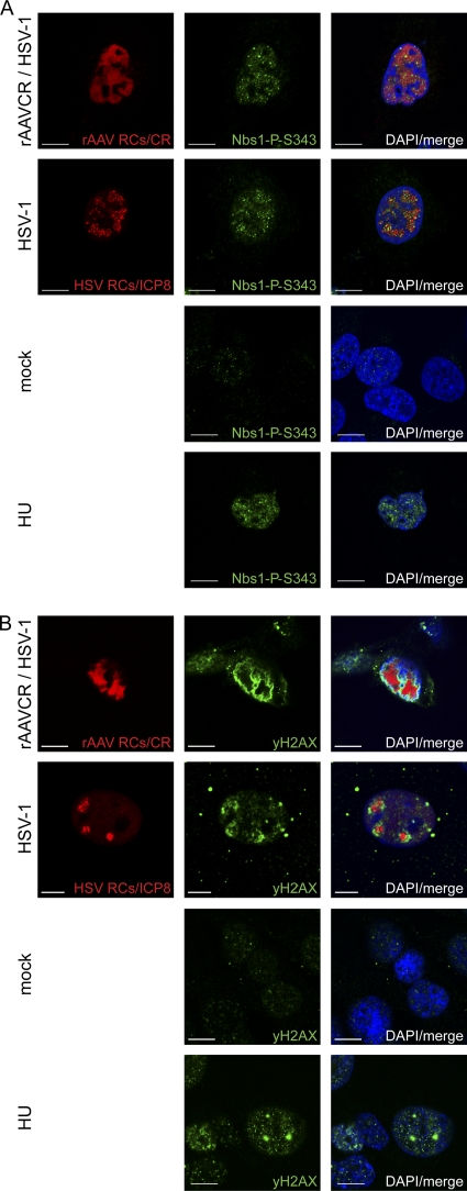
Fig 2.
Activation of primary DDR proteins. MO59J Fus1 cells were mock infected, infected with HSV-1 (MOI, 1.5), or coinfected with rAAVCR (MOI, 250) and HSV-1 (MOI, 1.5). After 24 h, cells were fixed and processed for IF analysis. rAAVCR RCs (AAV RCs) were visualized by binding of the rAAVCR-encoded mCherry-Rep68/78 fusion protein (CR) to AAV DNA (red). HSV-1 RCs were visualized with a primary antibody specific for the HSV-1 major DNA binding protein ICP8 and an AF594-labeled secondary antibody (red). Cells treated with HU (3 mM) served as a DDR control. To identify phosphorylated Nbs1 (A) and H2AX (B), cells were stained with antibodies specific for Nbs1-P-S343 or H2AX-P-S139 (γH2AX) and an FITC-labeled secondary antibody (green). DAPI was used to stain cellular DNA. Images were taken using a CLSM and represent a single optical z slice of the nuclei. Scale bars, 10 μm.

