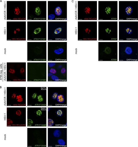
Fig 3.
Recruitment of ATM-P-S1981, ATR-P-S428, and ATRIP into HSV-1 and AAV2 RCs. MO59J Fus1 cells were infected and processed for IF, and viral RCs (red) were visualized as described in the legend to Fig. 2. Cells were stained with an antibody specific for ATM-P-S1981 (A), ATR-P-S428 (B), or ATRIP (C) and an FITC-labeled secondary antibody (green). DAPI was used to stain cell nuclei. In panel A, cells deficient for ATM (AT-22 IJE T) served as a control for the phosphospecific ATM antibody. In panel B, the percentage of ATR-P-S428 in viral RCs is indicated. Scale bars, 10 μm.

