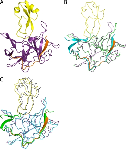
Fig 4.
DENV-3 protease-aprotinin structure and structural comparison with WNV protease-aprotinin and DENV-3 protease-Bz-nKRR-H structures. (A) Overview of the DENV-3 protease (NS3, purple; NS2B, orange) bound to aprotinin (yellow) shown in cartoon representation. (B) Alignment of the DENV-3 protease structures in complex with aprotinin (colored as in panel A) and in complex with Bz-nKRR-H (colored as in Fig. 1A). NS3 is shown in ribbon representation for clarity, with the rest in cartoon representation. (C) Comparison of the structures of DENV-3 protease-aprotinin (colored as in A) and WNV protease-aprotinin (NS3, blue; NS2B, green; aprotinin, gray), aligned on the Cα atoms of the protease only. NS2B is shown in cartoon representation, with the rest as ribbons.

