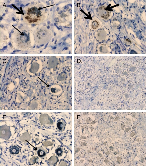
Fig 2.
Apparent VZV immunoreactivity and neuronal anti-type A reactivity are associated with the mouse ascites Golgi (MAG) staining artifact. DRG tissue sections were stained with anti-MAG antibody (1:64,000 dilution), which was generated in mice by pristane priming followed by intraperitoneal injection of hybridoma cells that were not from immunized mice. The DAB antibody-specific signal is brown (thick arrows), and the melanin counterstain is green (thin arrows). Panels A and B are sections from subject 4, who is blood type A, subtype A1; panels C and D are sections from subject 5, who is blood type A, subtype A2; and panels E and F are sections from subject 1, who is blood type O. Two representative images are shown for each condition. Magnification for panels on the left side, ×400. Magnification for panels on the right side, ×200.

