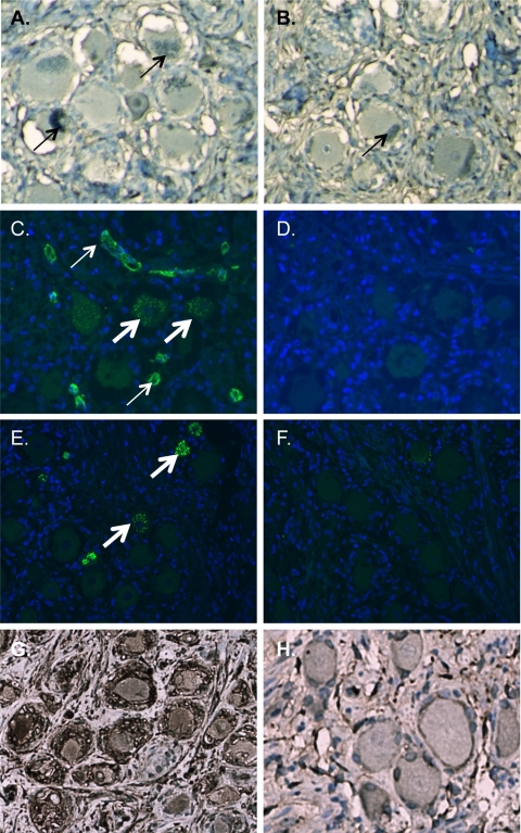
Fig 3.
Elimination of apparent VZV immunoreactivity in neurons using tissue culture-derived VZV monoclonal antibodies or by adsorption using blood type A erythrocytes. (A and B) Immunohistochemical staining of tissue sections from a blood type A subject with purified hybridoma supernatants from anti-IE62 (A) and anti-gE (B) clones at a 1:10 dilution eliminates immunoreactivity. Sections are counterstained with azure B, which stains neuromelanin green (thin arrows). (C to F) Immunofluorescent staining using a rabbit polyclonal antibody to IE62 and Alexa Fluor 488-conjugated anti-rabbit secondary antibody (C and D) or gE mouse ascites-derived monoclonal antibody and Alexa Fluor 488-conjugated anti-mouse secondary antibody (E and F). The primary antibody was adsorbed with blood type A erythrocytes (D and F) or unadsorbed (C and E). Thin arrows denote endothelial staining, and thick arrows denote staining of neuronal histo-blood group A determinants in Golgi zones. Sections for immunofluorescent staining were pretreated with Sudan black to eliminate the signal from neuronal pigments and counterstained with DAPI (4′,6-diamidino-2-phenylindole). (G and H) Immunohistochemical staining of VZV-infected DRG (G) and uninfected DRG (H) from a SCID mouse-human xenograft model. A VZV-specific signal with gE-mouse ascites-derived monoclonal antibody after adsorption with blood type A erythrocytes demonstrates that adsorption of endogenous anti-type A antibodies does not alter VZV-specific immunoreactivity. Sections are counterstained with hematoxylin.

