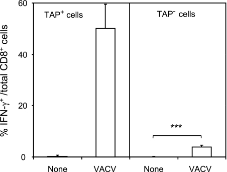
Fig 2.
Recognition of TAP+ and TAP− cell lines by VACV-specific CD8+ T lymphocytes. HLA-A*0201 TAP+ cells (RMA; left panel) and TAP− cells (RMA-S; right panel) were infected with VACV at a multiplicity of infection of 40 PFU/cell and analyzed by ICS for CD8+ T cell activation. The results are calculated as the means ± standard deviations (SD) of the results of three or four independent experiments. ***, P < 0.0001.

