. 2012 Jan;86(1):527–541. doi: 10.1128/JVI.05737-11

Fig 8.

Fig 8

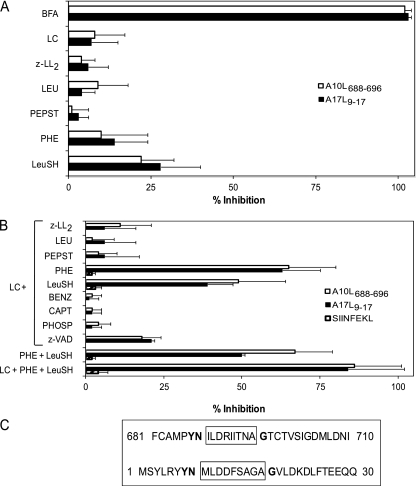
Recognition of target cells infected in the presence of inhibitors. (A and B) RMA-HLA-A*0201 target cells infected for 16 h with VACV or VACV-OVA257-264 at a multiplicity of infection of 40 PFU/cell were treated with the indicated inhibitors. An ICS assay was used to test for recognition by A10L688-696-specific (open bars), A17L9-17-specific (closed bars), or SIINFEKL-specific (hatched bars) CTLs. The percentages of specific inhibition obtained by the addition of the indicated single inhibitor (A) or mixture of two or three inhibitors (B) were calculated as follows:
%specificinhibition=100[(ICSVAVC+inhibitor)ICSwithoutinfection]ICSVACVICSwithoutinfection×100
where the data displayed represent the means ± SD of the results of 4 to 6 independent experiments. (C) The sequences of the residues neighboring the epitopes in A10L and A17L proteins. The sequence of each epitope is boxed. Residues that are identical in the two sequences are indicated with boldface characters. LC, lactacystin.