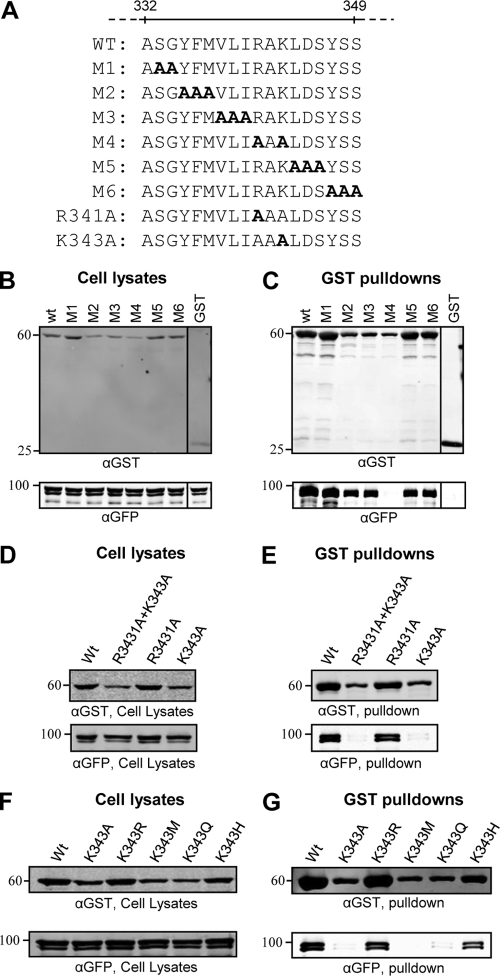
Fig 2.
Alanine scanning mutagenesis of VP16. (A) Amino acid sequence of the region spanning residues 332 to 349 of VP16 and mutants (M1 to M6) created in the context of GST-VP16(1–411). (B to G) 293T cells were cotransfected with GFP-VP1/2NT and indicated GST-VP16(1–411) mutants. Cell lysates were harvested at 48 h posttransfection and incubated with glutathione-Sepharose beads. Both cell lysates and bound protein complexes were separated by SDS-PAGE and analyzed by Western blotting with anti-GST and anti-GFP antibodies.

