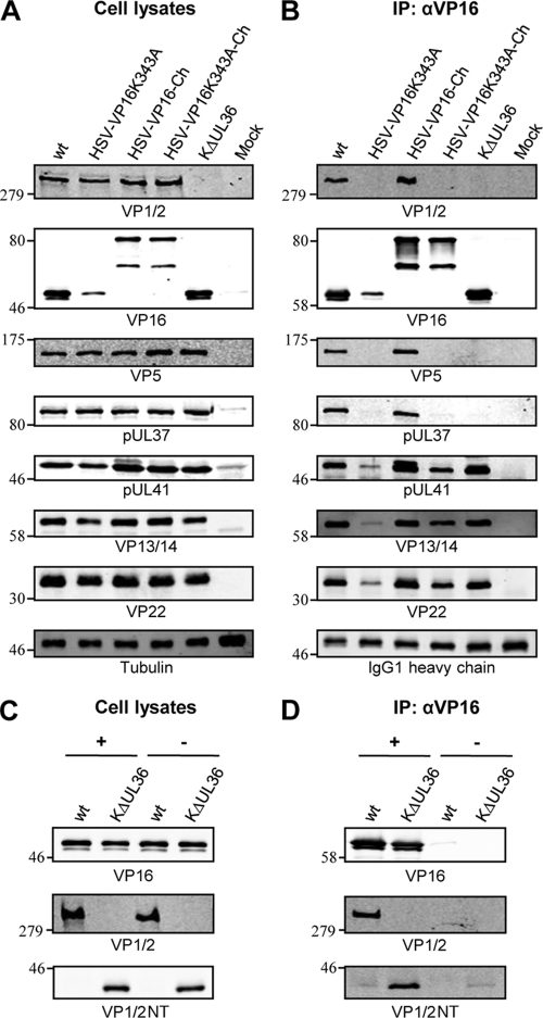
Fig 3.
K343A substitution in VP16 inhibits interaction with VP1/2 during infection. (A and C) HaCaT cells were infected with the indicated viruses, and cell lysates were harvested at 18 hpi. (B and D) Cell lysates were subjected to immunoprecipitation (IP) using an anti-VP16 (αVP16) antibody. Cell lysates (A and C) and bound protein complexes (B and D) were separated by SDS-PAGE and analyzed by Western blotting using the indicated antibodies. Tubulin and the heavy chain of the anti-VP16 antibody were included as loading controls.

