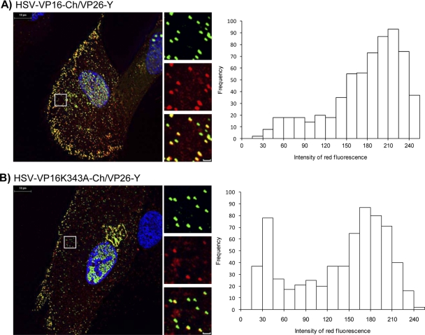
Fig 6.
Analysis of VP16-Ch and VP16(K343A)-Ch association with fluorescent capsids. Monolayers of HFF-hTERT cells grown on coverslips were infected with HSV-VP16-Ch/VP26-Y (A) or HSV-VP16(K343A)-Ch/VP26-Y (B). Cells were fixed and analyzed by fluorescence microscopy. Representative cells on right are shown with higher magnification (boxed area) slown along the right side of each large image. Scale bar of insets, 1 μm. Histograms at left show red fluorescence intensities of cytoplasmic EYFP-labeled particles.

