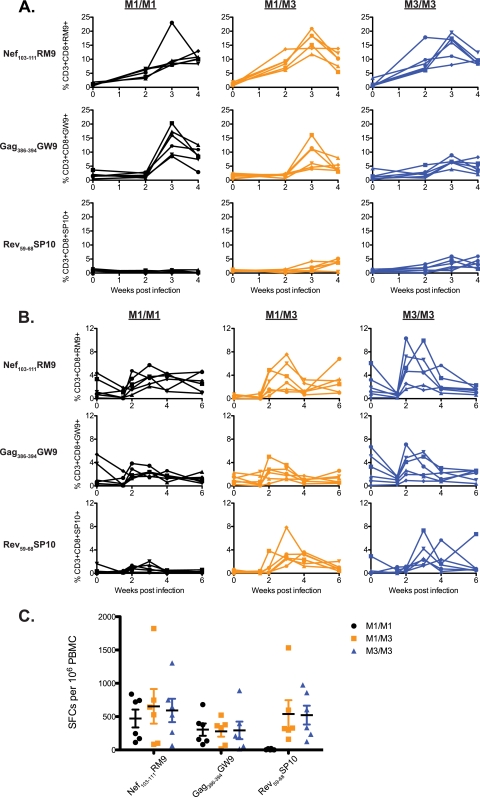
Fig 1.
Acute immunodominant CD8+ T cell responses in cells isolated from BAL fluid and peripheral blood samples. (A) MHC-peptide tetramers representing three SIVmac239-derived epitopes (Nef103-111RM9, Gag386-394GW9, and Rev59-68SP10) were used to quantify the percentages of antigen-specific CD3+ CD8+ T cells in BAL fluid samples at 0, 2, 3, and 4 weeks after SIVmac239 infection. (B) The same tetramers were used to quantify the percentages of antigen-specific CD3+ CD8+ T cells at 0, 1.5, 2, 3, 4, and 6 weeks after infection in peripheral blood samples. For panels A and B, six animals with each MHC genotype were followed: M1/M1 MCMs (black), M1/M3 MCMs (orange), and M3/M3 MCM (blue). Each shape represents a different animal. (C) T cell responses targeting the three SIV-derived peptide epitopes were measured by an IFN-γ ELISPOT assay 3 weeks after SIVmac239 infection. The average numbers of spot-forming colonies (SFCs) per 106 PBMC and the standard errors are shown. Six animals with each MHC genotype are shown as indicated in the legend.

